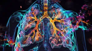
How Nuclear Medicine Plays a Role in Early Detection of Lung Diseases

Early detection is pivotal for effectively managing and treating lung diseases. Nuclear medicine, a highly advanced field of medical imaging, has emerged as a valuable tool for identifying lung conditions at an early stage. Using cutting-edge technology, this medical advancement provides detailed insights into lung function and abnormalities that may not be visible with conventional imaging techniques.
Understanding Nuclear Medicine
Nuclear medicine uses small amounts of radioactive material, known as radiotracers, to gather information about the body’s organs and tissues. Depending on the procedure, these radiotracers can be introduced into the body through injections, inhalation, or oral ingestion. Once inside the body, the radiotracers emit signals. Specialized imaging devices like PET or SPECT scanners capture these signals to create precise 3D representations of internal functions. This approach allows healthcare providers to go beyond structural imaging and assess the functionality of specific organs.
Common Lung Diseases and Challenges of Early Detection
Lung conditions, such as chronic obstructive pulmonary disease (COPD), lung cancer, pulmonary embolism, and interstitial lung diseases, often present with subtle or overlapping symptoms. Difficulty in distinguishing these conditions early contributes to delays in diagnosis and treatment. Symptoms like shortness of breath, chronic cough, or fatigue may be mistakenly attributed to less serious illnesses. Timely and accurate detection is necessary to prevent disease progression and improve outcomes. This advancement excels in providing functional analyses and identifying abnormalities before they manifest as physical symptoms.
How These Technologies Work in Lung Disease Detection
This advancement in medicine employs several techniques to aid in the early detection of lung conditions:
- Ventilation Perfusion (V/Q) Scans: V/Q scans are commonly used to evaluate blood flow and airflow in the lungs. They can help detect pulmonary embolisms—blockages in the lungs’ blood vessels that may go unnoticed with routine chest X-rays.
- Positron Emission Tomography (PET): PET scans use radiotracers to identify areas of abnormal metabolic activity within the lungs, often associated with tumors or inflammatory conditions. This technique is particularly valuable in detecting early-stage lung cancer.
- Lung Perfusion Imaging: Focused on assessing blood flow through the lungs, this imaging procedure is instrumental in diagnosing conditions like pulmonary hypertension and other circulation-related lung diseases.
Advantages in Early Diagnosis
The use of nuclear medicine in diagnosing lung diseases has several significant advantages:
- Functional Imaging Capability: Unlike X-rays or CT scans, which primarily provide structural views, functional imaging provides a more beneficial perspective for identifying issues like reduced airflow or blood supply.
- High Sensitivity: Nuclear medicine can detect even minor abnormalities at a molecular level, allowing it to identify potential problems long before they become apparent in traditional imaging methods.
- Non-Invasive and Minimal Discomfort: Procedures are generally noninvasive and cause minimal discomfort, making them more accessible to a wide range of patients.
Revolutionizing Lung Disease Detection
Nuclear medicine is revolutionizing lung disease detection, offering functional insights and enabling early intervention. By leveraging its advanced imaging techniques, healthcare providers can identify conditions sooner, leading to better outcomes and more effective management. To learn more or explore how nuclear medicine could improve your healthcare, consult a medical imaging specialist or contact a diagnostic radiology center near you.


